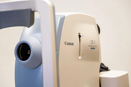
FFA (Fundus Fluorescein Angiography) is an imaging technique that allows for detailed examination of the eye's blood vessels and retinal layers. This technology is used by our expert physicians at SEVGİGÖZ to assess intraocular circulation and diagnose many retinal diseases.
FFA is performed by injecting a contrast agent called fluorescein into the bloodstream. As fluorescein circulates through the vessels, it reaches the eye tissues and the retina is imaged with the help of a camera. This allows for clear visualization of retinal vessels, leaks, blockages, and other abnormalities.
One of the most common applications of FFA is the evaluation of retinal diseases such as diabetic retinopathy, retinal vascular occlusions, and macular degeneration. FFA images play a critical role in determining the type and progression of the disease and provide important information for treatment planning.
The use of FFA has also become widespread in preoperative evaluation and surgical planning for retinal surgery. Especially in cases such as retinal detachment or retinal tears, FFA images are important for enhancing surgical success and improving postoperative outcomes.


