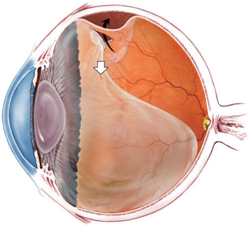
What is Retinal Detachment?
Retinal detachment is the separation of the layer of blood vessels beneath the light-sensitive nerve layer of the retina, which contains photoreceptors responsible for vision, from the retina. It occurs when the gel-like fluid inside the eye called vitreous leaks under the retina due to a tear or hole in the retina.What are the Risk Factors for Retinal Detachment?
Risk factors for retinal detachment include high myopia (over 6 diopters), family history, presence of retinal tears or detachment in the other eye, eye trauma, previous intraocular surgeries, and degenerative lesions that pose a risk of detachment.What are the Symptoms of Retinal Detachment?
Symptoms of retinal detachment may include flashes of light, light sensitivity, blurred vision, black spots, floaters, and loss of vision in the detached retina area.How is Retinal Detachment Diagnosed?
Retinal detachment can generally be detected through a detailed fundus examination with a biomicroscope after applying pupil-dilating drops. A detached retina appears swollen and white, and it undulates with eye movements.Retinal detachment can also be visualized using a Ocular Ultrasound (USG) device.
How is Retinal Detachment Treated?
If there are retinal tears without detachment, laser treatment with argon laser can be performed to seal the tear and prevent progression. The procedure is performed under topical anesthesia in the outpatient setting.If detachment has occurred, surgical treatment is determined based on the size and location of the detachment.
- Pneumatic Retinopexy: This method involves using gas to flatten the detached retina. It is suitable for small detachments in specific locations (upper quadrants). After injecting expanding gas into the eye, positioning is adjusted depending on the location of the tear.
- Scleral Buckling Surgery: In this procedure, a silicone band is placed around the eye to indent the sclera.
- Vitrectomy Surgery: This involves removing the vitreous fluid and applying laser around the tear. After surgery, a tamponade substance such as air, gas, or silicone is injected into the eye. If gas is injected into the eye, it is prohibited to travel to high altitudes or fly until the gas dissipates completely due to its expansion properties in response to atmospheric pressure.
The possibility of re-tear and detachment development in the future is always present.


