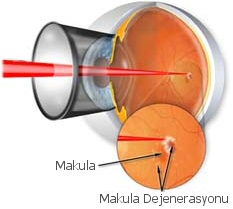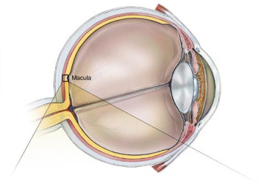
What is macular degeneration?
The center of the retina layer located on the back wall of the eyeball is called the "Macula." The macula region plays an important role in seeing objects clearly and in detail when looked straight ahead. Degeneration caused by the progressive loss of photoreceptor cells in this region is called Macular Degeneration and is commonly known as "Yellow Spot Disease." It is one of the most important causes of vision loss in older age.What are the risk factors for macular degeneration?
The most significant risk factor is advancing age, but other known risk factors include female gender, fair skin, colored eye structure, smoking, family history, genetic predisposition, and prolonged exposure to unprotected sunlight.How is macular degeneration identified?
In the early stages of macular degeneration, there are no symptoms. However, as the disease progresses, symptoms such as decreased central vision, seeing dark spots in the center of the field of vision, fading colors, distorted or wavy vision, and seeing floaters in the eyes often occur.What are the types of macular degeneration?
Macular degeneration is classified into two main types. Distinguishing between these two different conditions is important in making the diagnosis and planning treatment.Dry type:
The disease is mostly seen in this type (85-90%). It develops due to aging and progresses slowly. Vision loss is less frequent and occurs over a long period. It occurs due to thinning of the macula layer or accumulation of a yellow substance called drusen on the macula.Wet type:
It constitutes about 10-15% of macular degeneration cases. It occurs due to the formation of new blood vessels in the macula region, which causes leakage of blood and fluids from these vessels, disrupting the function of the cells responsible for vision. It leads to rapid and high rates of vision loss.Although approximately 80% of patients have the dry type, about 90% of central vision losses are caused by the wet type of macular degeneration.
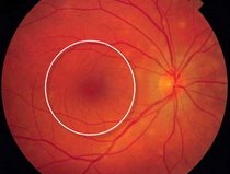
Normal Macula
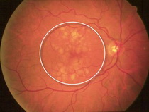
Dry Type Macular Degeneration
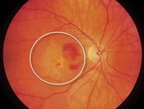
Wet Type Macular Degeneration
How is macular degeneration diagnosed?
- A detailed examination of the dilated fundus using a biomicroscope can detect the presence of specific findings such as drusen, bleeding, and exudation associated with the disease.
- Imaging methods such as Fundus Fluorescein Angiography (FFA) and Optical Coherence Tomography (OCT) can help detect the disease early.
- OCT is an important imaging method for understanding the progression of the disease and evaluating the response to treatment. It scans the central vision in thin sections using laser technology, providing results within a short period.
- FFA is a non-radiation angiography that involves injecting a contrast agent into the patient's forearm to better examine the retinal vascular structure.
How is macular degeneration treated?
Early diagnosis and treatment of macular degeneration can stop vision loss and slow the progression of the disease in many patients. Treatment is planned according to the subtype of the disease.The first step in treatment is regular doctor follow-up. Patients can also monitor themselves at home using a grid chart called the Amsler Grid test.
Avoiding smoking, exercising, avoiding direct exposure to sunlight, controlling cardiovascular diseases such as hypertension, consuming a diet rich in antioxidants from colorful fruits and vegetables, and consuming foods rich in Omega-3 fatty acids are general preventive measures.
In these patients, dietary supplementation with antioxidants such as Lutein, Zeaxanthin, vitamins A, C, E, and minerals such as Copper and Zinc, as well as vitamin supplements containing these substances, may slow the progression of the disease as recommended by the doctor.
New treatment methods are available for dry type macular degeneration. In recent years, a new treatment method called photobiomodulation (Valeda Laser) has been developed for dry type macular degeneration. It is an approved treatment using low-energy laser light to preserve the function of retinal cells. With this treatment, depending on the degree of degeneration, an increase in contrast sensitivity, an increase in visual acuity, and a decrease in accumulations called drusen can be achieved. The treatment is conducted in 9 sessions within a 3-week period, with each session lasting about 5 minutes. This treatment has no side effects.
Early intervention is necessary for the treatment of wet type macular degeneration.
Currently, there are three accepted types of treatment methods. These are Photodynamic Therapy, intravitreal injections of anti-VEGF drugs, and Laser Photocoagulation in selected cases. Anti-VEGF injections have surpassed the other two treatment options.
- Treatment known as Photodynamic Therapy is conducted in two stages. In the first stage, a special drug activated by light is administered to the patient intravenously, and the drug accumulates in the diseased tissue within 10 minutes. In the second stage, the drug is activated using a special non-burning laser applied externally, and the vascular mass in the macula layer is eliminated.
- Intravitreal injections of Anti-VEGF drugs are currently the most commonly used treatment option. Anti-VEGF drugs prevent the formation of vascular masses and promote their regression. They are administered into the vitreous cavity, the posterior chamber of the eye. Although there are various application regimens for these drugs, treatment continues based on OCT findings after an initial loading dose for 3 months.
- In selected cases, direct laser treatment may also be effective for leaking lesions located away from the macular center where new vessels are present.
